ABSTRACT
Objective: To compare the outcomes of thoracotomy and video-assisted thoracoscopic surgery (VATS) in the treatment of recurrence of primary spontaneous pneumothorax. Methods: Medical records of patients presenting recurrence of primary spontaneous pneumothorax were retrospectively reviewed. Patients were divided into two groups: those who underwent conservative thoracotomy (n = 53, thoracotomy group); and those who underwent VATS (n = 47, VATS group). Results: Although there were no deaths in either group and the length of hospital stays was similar between the two, there was greater morbidity in the thoracotomy group. Patients in the thoracotomy group required more pain medication for longer periods than did those in the VATS group (p < 0.05). In the thoracotomy group, the rate of recurrence was 3%. Pain was classified as insignificant at one month after the operation by 68% of patients in the VATS group and by only 21% of those in the thoracotomy group (p < 0.05). At three years after the surgical procedure, 97% of the VATS group patients considered themselves completely recovered from the operation, compared with only 79% in the thoracotomy group (p < 0.05). Chronic or intermittent pain, requiring the use of analgesics more than once a month, was experienced by 90% of the thoracotomy group patients and 3% of the VATS group patients. In addition, 13% of the thoracotomy group patients required clinical pain management. Conclusions: We recommend VATS as the first-line surgical treatment for patients with recurrent primary spontaneous pneumothorax.
Keywords:
Thoracoscopy; Thoracic surgery, video-assisted; Recurrence; Pneumothorax.
RESUMO
Objetivo: Comparar los resultados de la toracotomía con la video-assisted thoracoscopic surgery (VATS, cirugía torácica video-asistida) en el tratamiento de las recurrencias del neumotórax espontáneo primario. Métodos: Se revisaron los expedientes clínicos de los pacientes con neumotórax primario recurrente dividiéndose en dos grupos: pacientes sometidos a toracotomía (n = 53, grupo toracotomía) y pacientes sometidos a VATS (n = 47, grupo VATS). Resultados: La morbilidad fue mayor en el grupo A. Sin mortalidad en ninguno de los dos grupos. La duración de la hospitalización fue similar. Los pacientes del grupo toracotomía necesitaron más dosis de narcóticos durante períodos más largos de tiempo que los del grupo VATS (p < 0.05). Se presentó una recurrencia en el grupo toracotomía (3%). El 68% de los pacientes del grupo VATS y el 21% del grupo toracotomía clasificaron su dolor como insignificante transcurrido un mes de la cirugía (p < 0.05). A los tres años, el 97% de los sometidos a VATS se consideraron totalmente recuperados comparado con el 79% del grupo de toracotomía (p < 0.05). El 90% del grupo toracotomía y el 3% del grupo VATS se quejaban de dolor necesitando analgésicos por más de una vez al mes, 13% de los sometidos a toracotomía requirieron la asistencia de la clínica del dolor. Conclusiones: La VATS es la primera elección en el tratamiento de la recidiva del neumotórax espontáneo primario.
Palavras-chave:
Toracoscopía; Cirugía torácica asistida por video; Recurrencia; Neumotórax.
IntroductionThe most common cause of primary spontaneous pneumothorax (PSP) is the rupture of subpleural bullae or blebs located in the apex of the upper lobes, posteromedial border, or, more rarely, in the Fowler segment.(1,2) The optimal treatment of this type of benign lesions has been much debated.(3) Although conservative management through rest, needle aspiration or the placement of a drainage tube (thoracostomy) is safe, the rate of recurrence is high, ranging from 20% to 60%,(3,4) and thoracotomy is subsequently required.(5)
The lesions can be resected, and mechanical pleural abrasion or partial apical pleurectomy can be easily performed using less invasive approaches and incisions with better aesthetic results, such as axillary incision or mini-thoracotomy through the auscultatory triangle.(5,6) Despite the use of these limited incisions, many patients require a much longer recovery period.
Spontaneous pneumothorax can be effectively treated by thoracoscopy.(7) The indications for the intervention in cases of pneumothorax due to rupture of bullae or bullous emphysema are the same as those for performing the open procedure. These include recurrence or persistence (air leak for more than seven days) and a giant bulla that compresses the adjacent lung parenchyma, impairing its function.(8)
Surgical management depends on the etiology of the pneumothorax. If it is caused by the rupture of bullae, typically located in the apex of the upper lung lobes, treatment is simple.
If, however, it is due to bullous emphysema, treatment, even treatment using the conventional approach, can be quite complex.(8,9)
Thoracoscopy is not a novel procedure; it was first reported in 1910 by Jacobaeus,(10) who performed it using a Nitze cystoscope for intrapleural pneumonolysis due to TB and, in 1925, reported the outcomes in 120 patients. The method underwent significant advances based on the findings of Rodgers, who described the use of thoracoscopy in therapeutic procedures.(10)
Between 1993 and 2007, many surgeons have reported their experiences with video-assisted thoracoscopic surgery (VATS) in the treatment of PSP recurrence.(7,8,11) In those studies, VATS is compared with conventional thoracotomy by analyzing the early post-operative period, and the results were favorable.(9,10) Long-term follow-up evaluations were performed in few of those investigations. Those studies mainly focus on analyzing recurrence rates and pulmonary function.(11,12)
The objective of the present study was to compare the outcomes of VATS with those of standard thoracotomy, evaluating them in the short and long terms, with emphasis on the perspective of patients regarding quality of life and degree of satisfaction.
MethodsThe present study was approved by the Ethics Committee of and conducted in accordance with the guidelines for research involving human beings established by the Bolivarian Republic of Venezuela Ministry of Health and Social Assistance.
Clinical histories were reviewed for a population of 100 patients who underwent surgical treatment due to PSP recurrence between February of 1993 and July of 1999 at the Thoracic Surgery Division of the First Department of General Surgery of the Miquel Pérez Carreño University Hospital of the Venezuela Central University School of Medicine.
Patients were monitored for a mean follow-up period of 38 months (range, 30-53 months).
Inclusion criteriaThe inclusion criteria were as follows: medical history (hospital stay and office visit) being completely documented; being in good health; having no underlying lung disease (secondary pneumothorax); not having previously undergone thoracic surgery; and being between 21 and 55 years of age.
The variables studied included gender, smoking habits, type of intervention, intra-operative findings, intra-operative difficulties, type of lesion and location, duration of the intervention in minutes, complications, duration of thoracic drainage tube use, pain frequency/intensity as measured using a visual analog scale, dose of meperidine required after the removal of the epidural catheter and length of hospital stay.
In the office visit, the clinical and radiological documentation was registered, as were short- and long-term follow-up data, including pain frequency/intensity (0-5), need for analgesics or clinical pain management, return to normal activities (school, work, sports, etc.), episodes of recurrence, pulmonary function tests, evaluation of the cosmetic results, quality of life and degree of satisfaction with the procedure as evaluated using the Rensis Likert scale(13): strongly agree, agree, disagree or strongly disagree.
Exclusion criteriaThe exclusion criteria were as follows: being older than 55 years of age; medical history being incompletely documented regarding age, gender, smoking habits and side of the intervention; and being lost to long-term follow-up. Based on these criteria, a total of 20 patients were excluded. Of the remaining 80 patients, 45 (55.84%) had undergone conventional surgery and 35 (44.16%) had undergone VATS. Pain intensity in the post-operative period was measured using a visual analog scale. Patients were instructed to draw a vertical line crossing a horizontal line numbered from 0 to 5 (0 = absence of pain; 1 = occasional pain or discomfort; 2 = use of analgesics; 3 = frequent use of nonopioid analgesics; 4 = frequent use of opioids; and 5 = severe pain requiring clinical pain management) in order to represent the intensity of their pain.(13)
In the evaluation of the cosmetic results, patients were instructed to express their satisfaction regarding their scar using a 5-point scale (1 = very dissatisfied, 2 = dissatisfied, 3 = moderately satisfied, 4 = satisfied and 5 = very satisfied).(13)
Patients were asked to indicate the time elapsed between the intervention and the complete normalization of their activities: 1) less than one month; 2) more than three months; 3) more than 6 months; 4) more than one year; and 5) no complete recovery as yet.
The study population comprised 100 patients, who were divided into two groups based on the technique used.
Thoracotomy groupMini-thoracotomy was performed under general anesthesia using selective intubation with a double-lumen tube (Carlens tube).(12) Patients were positioned in the lateral decubitus position (contralateral hemithorax-sane side) on the surgical table. Limited axillary thoracotomy was performed with an incision of approximately 10 cm in length, and the planes were separated without sectioning the muscles. Blebs (single or multiple) were identified in the apex of the upper lobes and resected using an endoscopic linear cutting stapler (Endo-GIA; US Surgical Corp, Norwalk, CT, USA), three intercalated rows of titanium staples being placed on each side. Subsequently, mechanical abrasion of both pleural surfaces was performed. At the end of the procedure, a 36-F chest tube, with the tip facing the apex, was inserted for water-sealed thoracic drainage. The skin was sutured using interrupted intradermal sutures. Patients were extubated in the operating room and taken to the recovery room. Subsequently, patients were taken to the intensive care unit, where they remained for 48 h.
VATS groupThe position, preparation and surgical procedure were similar to those of conventional surgery. Combined (general and epidural) anesthesia was administered via a double-lumen Carlens tube. The video equipment (Karl Storz, Tuttingen, Germany) was positioned on either side of the head of the patient. The lung on the lesion side was collapsed, and a mini-incision was made in the fifth or sixth intercostal space on the posterior axillary line.(12) A trocar (10-mm endoscopic, 30 degrees, frontal view; connected to the camera), was placed (blind insertion) in the seventh or eighth intercostal space between the midaxillary line and the posterior axillary line. Two additional trocars (12-mm and 10-mm, respectively) were placed in the fourth intercostal space: the first at the posterior port and the second at the anterior port. The blebs were resected using the same type of instrument. The mechanical abrasion of both pleural serosae was performed in a manner similar to that of conventional surgery performed in recent years.
Post-operative carePatients were extubated in the operating room and observed for 2 h in the recovery room. During this interval, the chest drain was connected to a low-pressure suction system (−5 cmH2O) in order to reduce the residual pleural space and prevent the accumulation of blood clots. Subsequently, the chest drain was placed on water seal without applying negative pressure. The post-operative analgesia included the combination of continuous epidural anesthesia and the administration of nonsteroidal anti-inflammatory drugs. At the end of the second day, the epidural catheter was removed. Oral tramadol (200 mg/day) and dipyrone (4 g/day) were given.
Intramuscular meperidine was or was not prescribed based on the results of the evaluation of pain intensity and the needs of patients. If, at 24 h after the intervention, there was complete reexpansion of the lung, demonstrated by clinical and radiological evaluation, and no air leaks, the chest tube was removed. In the present study, prophylactic antibiotic therapy was not used, although antithrombotic agents were used routinely.
Patients were discharged on the following day with a prescription for continuation of the oral treatment, as well as for oxycodone if indicated.
Data were encoded and transcribed onto a matrix using the Statistical Package for the Social Sciences program, version 13 (SPSS Inc., Chicago, IL, USA). The descriptive analysis of the data included range (minimum and maximum values) and mean ± SD for continuous variables (age, surgical time, length of hospital stays and dose of meperidine), as well as the t-test and chi-square test for categorical variables, with values of p < 0.05 being considered significant.(14-16)
ResultsOf the population of 100 patients studied between 1984 and 1999, 53 underwent conventional thoracotomy, whereas 47 underwent minimally invasive VATS techniques (ratio, 1:1.12; proportion, 0.55).
Thirteen patients-9 in the thoracotomy group and 4 in the VATS group-were lost to long-term follow-up.
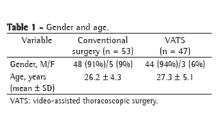
Clinical history was incomplete in 5% of the cases. Table 1 presents the demographic data (age and gender). More than half of the patients in both groups (64% and 65%, respectively) were heavy smokers, with a mean tobacco intake of more than 20 cigarettes per day.

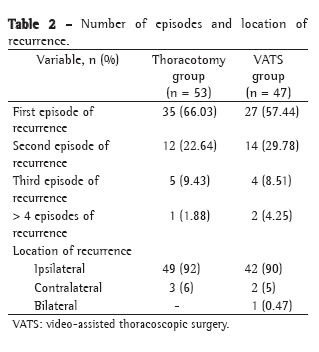
Table 2 shows the number of episodes of recurrence and their location.
The relationship between effort and recurrence was not significant in either group-93% in the thoracotomy group and 92% in the VATS group (p < 0.05).
Involvement of the right hemithorax was more common in both groups (57% and 60%, respectively). There was only one patient (0.47%) who underwent VATS due to bilateral recurrence.

Single or multiple blebs or bullae were seen in all of the cases. The site most commonly affected was the apical segment of the upper lobes, although the Fowler segment was occasionally affected (in 2%).
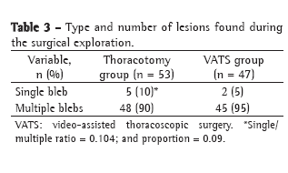
In both groups, patients in whom the number of episodes of recurrence was higher (3 or 4) presented intra-operative difficulties, which were caused by the presence of dense, fibrous connective pleuropulmonary adherences that increased surgical time, although the difference was not statistically significant. Table 3 shows the number of bullae found.

Mean intra-operative bleeding, measured in cubic centimeters, was greater in the open thoracotomy group patients (850 ± 50 cc) than in the VATS group patients (165 ± 25 cc; p < 0.05).
The mean surgical time in the thoracotomy group was 76 ± 15.6 min, compared with 52 ± 12.4 min in the VATS group (p = 0.05).
In the VATS group, there was no need to convert any of the interventions to conventional surgery.
All 53 patients in the conventional thoracotomy group were taken to the intensive care unit immediately after recovery in the postanesthesia care unit, compared with only 2 (4.25%) of the 47 patients in the VATS group.
The duration of chest tube drainage and hospital stays were shorter in the VATS group than in the conventional surgery group (p < 0.05). The epidural catheter was removed earlier in the VATS group than in the thoracotomy group (post-operative day 1 vs. post-operative day 5; p < 0.05).
The dose of meperidine required immediately after the epidural catheter had been removed was higher among the patients in the thoracotomy group than among those in the VATS group (365 ± 38 vs. 60 ± 18 mg/day; p < 0.05).
At hospital discharge, 81% of the thoracotomy group patients and 6% of the VATS group patients required analgesia with oral opioids (p < 0.05). Table 4 presents the short-term results.

In the thoracotomy group, recurrence was observed in 4 patients, 3 of whom were treated using chest tube drainage and one of whom required a second intervention.
Multiple bullae constituted the most common finding in both groups (in 90% and 95% of the patients, respectively). Single bullae were present in 10% of the thoracotomy group patients and in 5% of the VATS group patients.
Prolonged air leaks were more common in the VATS group (4%).
The dose of meperidine required to control pain, in milligrams per day, was higher in the conventional thoracotomy group patients (295 ± 48 mg/day) than in the VATS group patients (60 ± 18 mg/day; p < 0.05). In addition, the use of opioids at discharge, as well as at one week, two weeks and one month after discharge, was greater in the thoracotomy group than in the VATS group (81%, 81%, 52% and 26% vs. 6%, 8%, 4% and 0%, respectively; p < 0.05). At one month after discharge, 42% of the thoracotomy group patients reported pain, albeit mild.
Table 5 compares the two study groups in terms of the long-term results for the different variables.

Complete recovery was achieved in 69% of the thoracotomy group patients, compared with 99% of the VATS group patients (p < 0.05).
At three years after the intervention, the VATS group patients expressed greater satisfaction with the procedure, including quality of life and cosmetic results, than did those in the conventional surgery group (p < 0.05).
DiscussionIn 1956, the surgical treatment for recurrence of PSP was described.(17) Since then, many articles on the theme, evaluating various treatment methods, have been published.(18-21)
Limited axillary thoracotomy has been compared with mini-thoracotomy through the auscultatory triangle,(5,6) and partial pleurectomy has been balanced against pleural abrasion with or without bullectomy.(22-24)
Many authors have reported low morbidity rates and practically no mortality, as was the case in our study. The mean rate of PSP recurrence is below 5%.(25)
In 1990, some authors were the first to describe the use of VATS in the treatment of PSP recurrence.(22) Subsequently, there have been various publications involving large samples of patients treated by VATS and in which, as in our study, the safety of the procedure, percentage of conversion, length of hospital stay and short-term results were evaluated.(7,8,11,13) Those studies, similarly to ours, confirm the advantages of minimally invasive techniques, as does a comparative article,(23)
published in 1997, in which the authors showed that patients who undergo VATS report lower pain intensity, recover more rapidly and require shorter hospital stays than do those undergoing other types of surgery.
In other studies, in which long-term results are evaluated, the principal outcome measures were the rate of recurrence and the results of pulmonary function tests.(11,12,19,24) Very few investigations have evaluated, as we have in the present study, long-term follow-up by assessing, from the patient perspective, the frequency and intensity of pain, functional results, period of incapacity, quality of life and satisfaction with the procedure-these being the variables for which the greatest statistical difference has been found between minimally invasive techniques and conventional surgery.(25)
In the present study, which is similar to a study published in 2006,(25) we compared early and late results obtained in a group of patients treated through limited thoracotomy with those obtained in a group of patients treated through VATS.
The mean surgical time, from skin incision to placement of the drainage tube, was shorter in the patients who underwent VATS (52 ± 12.4 min) than in those who underwent conventional surgery (76 ± 15.6 min; p < 0.05).
The length of hospital stay was significantly shorter in the patients who underwent minimally invasive thoracic surgery than those who underwent open surgery (p < 0.05). The former suffered less intense pain and required lower doses of opioids for shorter periods, with a reduction in side effects (nausea and constipation). These differences confirm our hypothesis that, in the immediate post-operative period, the tolerance for VATS is better due to its minimally invasive character, with less intra-operative tissue trauma in comparison with that occurring during conventional thoracotomy.
The long-term differences found in the present investigation are even more pronounced and significant (Table 5). In our experience, PSP is a benign disease that most commonly affects young healthy individuals, primarily males, with a long life expectancy, having many years of physical activities, including work and sports, ahead of them. The psychological and economic consequences of prolonged pain and debility should be avoided as much as possible.(22)
At three years after the intervention, more than 25% of the thoracotomy group patients evaluated in the present study continued to receive pain medication, and only 63% considered themselves completely recovered from the operation.
Only 63% of the thoracotomy group patients were satisfied with the outcome of the surgical procedure, compared with 98% of those in the VATS group.
Based on these data, we conclude that VATS is the procedure of choice for the treatment of PSP recurrence. These conclusions are supported not only by the favorable results observed in the immediate post-operative period but also by the significantly better long-term results.
References 1. Torresini G, Vaccarili M, Divisi D, Crisci R. Is video-assisted thoracic surgery justified at first spontaneous pneumothorax? Eur J Cardiothorac Surg. 2001;20(1):42-5.
2. Chan P, Clarke P, Daniel FJ, Knight SR, Seevanayagam S. Efficacy study of video-assisted thoracoscopic surgery pleurodesis for spontaneous pneumothorax. Ann Thorac Surg. 2001;71(2):452-4.
3. Freixinet JL, Canalís E, Juliá G, Rodriguez P, Santana N, Rodriguez de Castro F. Axillary thoracotomy versus videothoracoscopy for the treatment of primary spontaneous pneumothorax. Ann Thorac Surg. 2004;78(2):417-20.
4. Sawada S, Watanabe Y, Moriyama S. Video-assisted thoracoscopic surgery for primary spontaneous pneumothorax: evaluation of indications and long-term outcome compared with conservative treatment and open thoracotomy. Chest. 2005;127(6):2226-30.
5. Ferraro P, Beauchamp G, Lord F, Emond C, Bastien E. Spontaneous primary and secondary pneumothorax: a 10-year study of management alternatives. Can J Surg. 1994;37(3):197-202.
6. Donahue DM, Wright CD, Viale G, Mathisen DJ. Resection of pulmonary blebs and pleurodesis for spontaneous pneumothorax. Chest. 1993;104(6):1767-9.
7. Janssen JP, van Mourik J, Cuesta Valentin M, Sutedja G, Gigengack K, Postmus PE. Treatment of patients with spontaneous pneumothorax during videothoracoscopy. Eur Respir J. 1994;7(7):1281-4.
8. Cardillo G, Facciolo F, Giunti R, Gasparri R, Lopergolo M, Orsetti R, et al. Videothoracoscopic treatment of primary spontaneous pneumothorax: a 6-year experience. Ann Thorac Surg. 2000;69(2):357-61; discussion 361-2.
9. Kim KH, Kim HK, Han JY, Kim JT, Won YS, Choi SS. Transaxillary minithoracotomy versus video-assisted thoracic surgery for spontaneous pneumothorax. Ann Thorac Surg. 1996;61(5):1510-2.
10. Rodgers BM, Champion JK, Wain JC. Modern Thoracoscopy. In: Arregui ME, Fitzgibbons Jr RJ, Katkhouda N, McKeman JB, Reich H, editors. Principles of laparoscopic surgery: basic and advanced techniques. New York: Springer-Verlag; 1995. p. 517-31.
11. Mouroux J, Elkaïm D, Padovani B, Myx A, Perrin C, Rotomondo C, et al. Video-assisted thoracoscopic treatment of spontaneous pneumothorax: technique and results of one hundred cases. J Thorac Cardiovasc Surg. 1996;112(2):385-91.
12. Krasna M, Mack MJ. Atlas of thoracoscopic surgery. St. Louis: Quality Medical Pub; 1994.
13. Sampieri RH, Collado CF, Lucio BP. Metodología de la investigación. México City: McGraw-Hill/Interamericana; 2003.
14. Ferran Aranaz M. SPSS para Windows: Análisis estadístico. Biblioteca profesional. Madrid: McGraw-Hill/Interamericana; 2001.
15. Chacín Lugo F. Diseño y análisis de experimentos para generar superficies de respuesta. Maracay (Venezuela): Universidad Central de Venezuela, Facultad de Agronomía; 2000.
16. Salama D. Estadística: Metodología y aplicaciones. 5th ed. Caracas: Editorial Principios; 2002.
17. Gaensler EA. Parietal pleurectomy for recurrent spontaneous pneumothorax. Surg Gynecol Obstet. 1956;102(3):293-308.
18. Bertrand PC, Regnard JF, Spaggiari L, Levi JF, Magdeleinat P, Guibert L, et al. Immediate and long-term results after surgical treatment of primary spontaneous pneumothorax by VATS. Ann Thorac Surg. 1996;61(6):1641-5.
19. Passlick B, Born C, Häussinger K, Thetter O. Efficiency of video-assisted thoracic surgery for primary and secondary spontaneous pneumothorax. Ann Thorac Surg. 1998;65(2):324-7.
20. Moores DWO. Pleurodesis by mechanical pleural abrasion for spontaneous pneumothorax. In: Deslaurieus J, Lacquet LK. editors. Thoracic Surgery: Surgical Management Of Pleural Diseases; International Trends In General Thoracic Surgery Volume 6. St Louis: Mosby; 1990. p 126-9.
21. Maggi G, Ardissone F, Oliaro A, Ruffini E, Cianci R. Pleural abrasion in the treatment of recurrent or persistent spontaneous pneumothorax. Results of 94 consecutive cases. Int Surg. 1992;77(2):99-101.
22. Levi JF, Kleinmann P, Riquet M, Debesse B. Percutaneous parietal pleurectomy for recurrent spontaneous pneumothorax. Lancet. 1990;336(8730):1577-8.
23. Jiménez-Merchán R, García-Díaz F, Arenas-Linares C, Girón-Arjona JC, Congregado-Loscertales M, Loscertales J. Comparative retrospective study of surgical treatment of spontaneous pneumothorax. Thoracotomy vs thoracoscopy. Surg Endosc. 1997;11(9):919-22.
24. Waller DA, Yoruk Y, Morritt GN, Forty J, Dark JH. Videothoracoscopy in the treatment of spontaneous pneumothorax: an initial experience. Ann R Coll Surg Engl. 1993;75(4):237-40.
25. Ben-Nun A, Soudack M, Best LA. Video-assisted thoracoscopic surgery for recurrent spontaneous pneumothorax: the long-term benefit. World J Surg. 2006;30(3):285-90.
About the authors
Jorge Ramón Lucena Olavarrieta
Full Professor. Central University of Venezuela, Caracas, Venezuela.
Pául Coronel
Instructor. Central University of Venezuela, Caracas, Venezuela.
Study carried out at the Central University of Venezuela, Caracas, Venezuela.
Correspondence to: Jorge Ramón Lucena Olavarrieta. Universidad Central de Venezuela, Cátedra de Técnica Quirúrgica, Instituto Anatómico José Izquierdo, oficina 213, Sucre, 1060, Caracas, Venezuela.
Telefax 58 021 2986-3458. E-mail: jorge_lucena@yahoo.com
Financial support: This study received financial support from the Council for Scientific and Human Development, Central University of Venezuela PI No 09-00-6197-2007.
Submitted: 18 February 2008. Accepted, after review: 1 July 2008.


