Ao Editor:A infecção por influenza é a mais comum infecção viral que causa doença respiratória. A pneumonia por influenza é a complicação mais séria da infecção pelo vírus da influenza e resulta em elevada mortalidade.(1) Embora os vírus da influenza sazonal sejam comumente detectados por meio de testes rápidos de antígeno com swabs de secreção de nasofaringe, raramente se isola um vírus da gripe suína (H1N1).(1) Portanto, a terapia anti-influenza deve ser iniciada empiricamente caso haja suspeita de pneumonia por influenza.
Sabe-se que o vírus da parainfluenza (VPI) causou doença semelhante à gripe durante as pandemias de influenza suína.(2) Relatou-se que o VPI 3 (VPI3) pode causar pneumonia em pacientes imunossuprimidos, tais como adultos que receberam transplantes.(3)
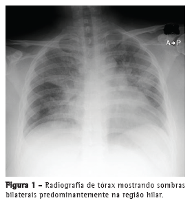
Relatamos um caso, tratado com sucesso, de pneumonia por VPI3 simulando pneumonia por influenza em uma paciente asmática de 31 anos de idade. A paciente apresentou febre alta (39.5°C), fadiga geral, dor articular sistêmica e anorexia durante dois dias antes de ser encaminhada a nosso centro médico. Era fumante e apresentava história de tabagismo (20 anos maço) e de asma brônquica (sem uso atual de medicação). A paciente também apresentava diabetes mellitus mal controlada e índice de massa corporal de 30 kg/m2. A radiografia de tórax revelou opacidades em vidro fosco difusas em ambos os pulmões (Figura 1). Exames laboratoriais revelaram reação inflamatória grave (proteína C reativa = 19,2 mg/dL e VHS = 83 mm/h). A paciente apresentou insuficiência respiratória grave e SpO2 de 80% em ar ambiente na primeira visita e passou a receber oxigenoterapia com ventilação não invasiva com pressão positiva. Devido à insuficiência respiratória grave, não foi realizada lavagem broncoalveolar. Embora o resultado de um teste rápido para detecção de antígeno de influenza tenha sido negativo, a paciente recebeu diagnóstico de pneumonia por influenza com base em sintomas semelhantes aos da gripe e em achados radiológicos, tais como opacidades difusas em vidro fosco (Figura 2).


A paciente passou a receber tratamento empírico com peramivir (600 mg/dia) durante 5 dias (para a infecção por influenza) associado a pulso de esteroide e eritromicina i.v. (1.000 mg/dia) durante 5 dias (para a insuficiência respiratória aguda). Sua função respiratória melhorou gradualmente, e a ventilação não invasiva com pressão positiva foi interrompida no 5º dia. A paciente passou então a receber prednisolona oral (80 mg/dia), e a dose foi sendo gradativamente reduzida uma vez a cada três dias, da seguinte maneira: para 40 mg/dia no 6º dia; para 30 mg/dia no 9º dia; para 15 mg/dia no 12º dia e suspensa no 15º dia. A paciente recebeu alta no 18º dia. Embora os resultados dos testes para anticorpos contra influenza A e B tivessem sido negativos, o resultado do teste para anticorpos contra VPI3 foi positivo. Estabeleceu-se o diagnóstico final de pneumonia por VPI3.
Os simuladores mais comuns de pneumonia por influenza suína (H1N1) são a doença dos legionários e a pneumonia por VPI3 humana (VPI3H) em adultos e a pneumonia causada por vírus sincicial respiratório ou metapneumovírus humano em crianças.(4) Sabe-se que os VPI humana tipos 1-3 são primordialmente patógenos respiratórios pediátricos e causa comum de laringotraqueobronquite (crupe) em crianças pequenas.(4) Em adultos, o VPI3H é reconhecido como causa de pneumonia adquirida na comunidade (PAC) em pacientes imunossuprimidos ou em pacientes que receberam transplantes. Entretanto, o VPI3H também pode se apresentar na forma de PAC viral em hospedeiros normais.(5) Durante uma pandemia de influenza, os clínicos devem considerar o diagnóstico de pneumonia por VPI3 caso os pacientes apresentem sintomas semelhantes aos da gripe e resultados negativos nos testes rápidos para detecção do antígeno do vírus da influenza.
Embora tenha havido relatos de casos tratados com sucesso,(2) o tratamento-padrão para pneumonia por VPI3 ainda não foi definido. Administramos pulsoterapia com esteroide (eritromicina i.v.) associada a medicação antigripe. O resultado foi que a função respiratória da paciente melhorou rapidamente. Relatou-se que macrolídeos podem ser eficazes contra inflamação grave, já que é evidente que reduzem a quimiotaxia de neutrófilos e a infiltração no epitélio respiratório, infrarregulando a expressão de moléculas de adesão e aumentando a apoptose de neutrófilos.(6,7) Além disso, estudos com animais demonstraram que altas doses de esteroides conseguem reduzir as lesões pulmonares e suprimir as citocinas de maneira eficaz, levando a uma interrupção da ativação de macrófagos.(6-8) É razoável supor que, no presente caso, a eritromicina teve um efeito imunomodulador em vez de um efeito antimicrobiano quando combinada com uma dose alta de prednisolona.
Os níveis plasmáticos de citocinas como IL-6 e IL-10 revelaram-se mais elevados em pacientes infectados pelo vírus da influenza A (H1N1) que morreram ou que apresentaram síndrome do desconforto respiratório agudo do que em pacientes com doença menos grave.(9) O mecanismo da inflamação pulmonar grave ainda não está claro. As adipocinas liberadas pelos adipócitos poderiam causar uma reação alérgica. Além disso, uma tempestade de citocinas poderia facilmente ocorrer em pacientes com doenças alérgicas como asma brônquica e doença reumática.(10) Por ser obesa e ter história de asma brônquica, nossa paciente apresentava risco de inflamação pulmonar grave.
Uma limitação de nosso relato é que os níveis plasmáticos de citocinas não foram determinados. Como marcadores inflamatórios como a proteína C reativa e a VHS estavam muito elevados, especulamos que tenha ocorrido uma tempestade de citocinas em nossa paciente.
Em suma, os médicos devem estar cientes de que outros patógenos podem causar PAC viral simulando pneumonia por influenza durante uma pandemia de influenza. Devem ser investigados mais casos a fim de definir o tratamento-padrão para pneumonia viral.
AgradecimentosAgradecemos ao Sr. John Wocher, Vice-Presidente Executivo e Diretor, Assuntos Internacionais/Serviços para Pacientes Internacionais, Kameda Medical Center, Kamogawa, Japão, a leitura crítica diligente e minuciosa de nosso manuscrito.
Nobuhiro Asai
Residente, Kameda Medical Center, Kamogawa, Japão
Yoshihiro Ohkuni
Chefe, Kameda Medical Center, Kamogawa, Japão
Norihiro Kaneko
Chefe, Kameda Medical Center, Kamogawa, Japão
Yasutaka Kawamura
Chefe do Departamento de Radiologia, Kameda Medical Center,
Kamogawa, Japão
Masahiro Aoshima
Chefe do Departamento de Pneumologia, Kameda Medical Center, Kamogawa, Japão
Referências1. Itoh Y, Shinya K, Kiso M, Watanabe T, Sakoda Y, Hatta M, et al. In vitro and in vivo characterization of new swine-origin H1N1 influenza viruses. Nature. 2009;460(7258):1021-5. PMid:19672242 PMCid:2748827.
2. Cunha BA, Corbett M, Mickail N. Human parainfluenza virus type 3 (HPIV 3) viral community-acquired pneumonia (CAP) mimicking swine influenza (H1N1) during the swine flu pandemic. Heart Lung. 2011;40(1):76-80. PMid:20888645. http://dx.doi.org/10.1016/j.hrtlng.2010.05.060
3. Whimbey E, Vartivarian SE, Champlin RE, Elting LS, Luna M, Bodey GP. Parainfluenza virus infection in adult bone marrow transplant recipients. Eur J Clin Microbiol Infect Dis. 1993;12(9):699-701. PMid:8243487. http://dx.doi.org/10.1007/BF02009383
4. Cunha BA, Pherez FM, Strollo S. Swine influenza (H1N1): diagnostic dilemmas early in the pandemic. Scand J Infect Dis. 2009;41(11-12):900-2. PMid:19922079. http://dx.doi.org/10.3109/00365540903222465
5. Marx A, Gary HE Jr, Marston BJ, Erdman DD, Breiman RF, Török TJ, et
al. Parainfluenza virus infection among adults hospitalized for lower respiratory tract infection. Clin Infect Dis. 1999;29(1):134-40. PMid:10433576. http://dx.doi.org/10.1086/520142
6. Amsden GW, Baird IM, Simon S, Treadway G. Efficacy and safety of azithromycin vs levofloxacin in the outpatient treatment of acute bacterial exacerbations of chronic bronchitis. Chest. 2003;123(3):772-7. PMid:12628877. http://dx.doi.org/10.1378/chest.123.3.772
7. Asai N, Ohkuni Y, Matsunuma R, Iwama K, Otsuka Y, Kawamura Y, et al. A case of novel swine influenza A (H1N1) pneumonia complicated with virus-associated hemophagocytic syndrome. J Infect Chemother. 2012. [Epub ahead of print]. http://dx.doi.org/10.1007/s10156-011-0366-3
8. Ottolini M, Blanco J, Porter D, Peterson L, Curtis S, Prince G. Combination anti-inflammatory and antiviral therapy of influenza in a cotton rat model. Pediatr Pulmonol. 2003;36(4):290-4. PMid:12950040. http://dx.doi.org/10.1002/ppul.10320
9. Writing Committee of the WHO Consultation on Clinical Aspects of Pandemic (H1N1) 2009 Influenza, Bautista E, Chotpitayasunondh T, Gao Z, Harper SA, Shaw M, et al. Clinical aspects of pandemic 2009 influenza A (H1N1) virus infection. N Engl J Med. 2010;362(18):1708-19. Erratum in: N Engl J Med. 2010;362(21):2039. PMid:20445182. http://dx.doi.org/10.1056/NEJMra1000449
10. Shore SA, Terry RD, Flynt L, Xu A, Hug C. Adiponectin attenuates allergen-induced airway inflammation and hyperresponsiveness in mice. J Allergy Clin Immunol. 2006;118(2):389-95. PMid:16890763. http://dx.doi.org/10.1016/j.jaci.2006.04.021


