ABSTRACT
Pulmonary sequestration is an uncommon condition that accounts for 0.5-6% of all pulmonary malformations and is typically diagnosed in childhood. Of the two forms of pulmonary sequestration, intralobar and extralobar, the latter is less frequently encountered. The current report describes the case of a 32-year-old female patient with chest and abdominal pain. Imaging (chest X-rays and computed tomography scans of the chest) revealed consolidation and pleural effusion. The initial thoracocentesis revealed hemothorax. Subsequent diagnostic video-assisted thoracoscopy revealed extralobar pulmonary sequestration. Consequently, the therapeutic decision was to make the conversion to thoracotomy in order to resect the lesion and safely ligate the intercostal vascular pedicle.
Keywords:
Bronchopulmonary sequestration; Hemothorax; Pulmonary infarction.
RESUMO
O sequestro pulmonar é uma malformação incomum, representando 0,5-6% de todas as malformações pulmonares, sendo geralmente diagnosticado na infância. Dos dois tipos de sequestro pulmonar, intralobar e extralobar, este último é o menos freqüente. O presente relato descreve o caso de um paciente do sexo feminino, de 32 anos, com quadro de dor toracoabdominal e achados de radiografia e TC de tórax revelando consolidação e derrame pleural. A conduta inicial com toracocentese evidenciou hemotórax. A seqüência diagnóstica através da videotoracoscopia permitiu o diagnóstico de sequestro extralobar e a consequente conduta de conversão para toracotomia para ressecção da lesão com ligadura segura do pedículo vascular intercostal.
Palavras-chave:
Sequestro broncopulmonar; Hemotórax; Infarto pulmonar.
IntroduçãoO sequestro pulmonar é uma doença incomum, com uma incidência de aproximadamente 1,5% das malformações pulmonares congênitas.(1-4)
Caracterizada pela aberrante nutrição arterial derivada da circulação sistêmica, o sequestro pulmonar divide-se em sequestro intralobar (SIL) e extralobar (SEL). Seu diagnóstico, na maioria dos casos, decorre de achados radiológicos ocasionais. No entanto, quando presentes, as manifestações clínicas são caracterizadas por pneumonias de repetição na infância, localizadas predominantemente no lobo inferior do pulmão esquerdo.(3,5)
Menos frequente do que o SIL, o SEL, por não apresentar qualquer contato com a árvore brônquica, raramente se manifesta com sintomas clínicos e está associado mais comumente com outras malformações congênitas. Apresentamos um caso de SEL que se manifestou com dor torácica e hemotórax, diagnosticado por videotoracoscopia, e integralmente ressecado através de toracotomia aberta.
Relato de casoPaciente feminino, 32 anos, branca, atendida na emergência de um hospital terciário, com quadro de dor intensa, de início súbito, na transição toracoabdominal, à direita, com 24 h de evolução. Apresentava-se inquieta, taquipnéica (frequência respiratória, 22 ciclos/min), temperatura axilar de 37,7°C. Exames laboratoriais gerais dentro dos limites da normalidade. Amamentando há nove meses. No atendimento inicial, foram aventadas e descartadas as hipóteses de tromboembolismo pulmonar e de cólica renal.
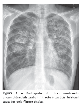
No entanto, a radiografia de tórax mostrava obliteração do seio costofrênico direito. O estudo tomográfico de tórax confirmou a presença de derrame pleural associado a uma área de consolidação (Figura 1). A avaliação do serviço de cirurgia torácica indicou a realização de toracocentese pela suspeita diagnóstica de derrame parapneumônico. A punção pleural revelou a presença inequívoca de um hemotórax. Mediante este achado, foi proposta a exploração do espaço pleural através de videotoracoscopia, que identificou hemotórax de moderadas proporções (aproximadamente 500 mL) junto à goteira paravertebral. Após a aspiração do conteúdo sangüíneo, foi possível a visualização de uma área de aspecto hepatizado, sem contato com o pulmão.

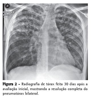
A lesão estava presa à coluna vertebral, no recesso costodiafragmático, na emergência do segmento costal junto à apófise transversa, por uma estrutura semelhante a um pedículo vascular. Decidiu-se pela conversão para toracotomia aberta para melhor abordagem deste pedículo e remoção da lesão (Figura 2).

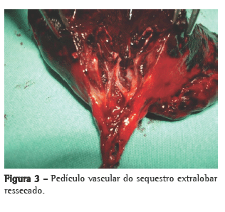
Os achados sugeriam fortemente a presença de um sequestro com torção e ruptura parcial do pedículo vascular e infarto do segmento pulmonar extralobar. A ressecção da lesão foi alcançada após a ligadura do pedículo vascular oriundo da artéria e veia intercostais. O segmento da veia intercostal anômala mostrava evidências de ruptura e colapso prévios (Figura 3), o que poderia explicar o sangramento. A cavidade pleural foi drenada com dreno tubular de calibre 28 French, retirado no segundo dia de pós-operatório.

A evolução cirúrgica transcorreu sem anormalidades, e a paciente recebeu alta no terceiro dia de pós-operatório.
No estudo anatomopatológico foi comprovado o diagnóstico de SEL com infarto caracterizado por necrose e hemorragia da lesão, secundário à rotação do pedículo vascular sobre o próprio eixo.
A possibilidade de que a gravidez recente tenha mobilizado a área da sequestro e induzido a rotação do pedículo foi considerada como a mais provável para explicar a apresentação deste caso.
DiscussãoO termo sequestro, que deriva do latim sequestare e significa separar, foi primariamente utilizado por Pryce, em 1946, quando ele descreveu um caso de SIL e despertou o interesse clínico pela entidade.(1,2)
Posteriormente, outros autores associaram anomalias pulmonares com anormalidades do suprimento sanguíneo, da via aérea e do desenvolvimento do intestino embriológico e do diafragma.(1-3)
Atualmente, as malformações do pulmão têm uma incidência estimada em 2,2-6,6%, sendo o sequestro pulmonar a segunda malformação mais freqüente (0,15-1,8%).(1) O sequestro pulmonar é definido como uma massa de tecido pulmonar não-funcionante que recebe suprimento sanguíneo de uma artéria sistêmica anômala e que não se comunica com a árvore brônquica normal, sendo dividido em SIL e SEL.(1-4,6,7) O SEL possui um revestimento pleural próprio, enquanto o SIL compartilha do mesmo revestimento pleural do pulmão normal.(3,7)
O SIL perfaz entre 75% e 85% dos achados de sequestro pulmonar(1,7) e é considerado uma doença adquirida, com episódios de infecções pulmonares recorrentes acometendo cerca de 50% dos pacientes até os 20 anos de idade.(1,5,6) Os lobos inferiores estão envolvidos em 98% dos casos e, mais comumente, à esquerda (55-60%), sempre acima do diafragma. A artéria que supre o sequestro deriva geralmente da aorta torácica, enquanto que a drenagem venosa tipicamente é realizada pelas veias pulmonares (95%).(5-7) Raramente, há associação com malformações congênitas, e não há predominância sexual na ocorrência do SIL.
O SEL é considerado uma doença congênita resultante de um ramo anormal do botão pulmonar que, em alguns casos, manteria a ligação original com o intestino, permitindo uma comunicação entre o sequestro e o sistema gastrointestinal.(7) Tipicamente é uma lesão assintomática, única, ovóide ou piramidal, que varia de 3 a 6 cm.(7) O diagnóstico de SEL é geralmente feito através de um achado ocasional em radiologia torácica de rotina ou incidentalmente durante uma laparotomia ou toracotomia
O hemitórax esquerdo é o mais acometido (65% dos casos), e o sítio principal do SEL é entre o lobo pulmonar inferior e o diafragma (63%), podendo também ser encontrado em topografia intra-abdominal (10-15%), mediastino anterior (8%) e mediastino posterior (6%).(3) Existe predominância do sexo masculino sobre o feminino, numa razão de 3-4:1.(3,6,7).
Em mais de 60% dos casos de SEL, coexistem outras anomalias congênitas, sendo a hérnia diafragmática a mais comum (16%).
Aproximadamente 25% dos casos possuem outra anormalidade pulmonar associada, como hipoplasia pulmonar, malformação adenomatóide cística e enfisema lobar congênito.(1,3,7)
Em mais de 80% dos casos, o suprimento arterial do SEL provém da aorta torácica ou abdominal. Em 15%, a artéria responsável pelo aporte sanguíneo advém de outra artéria sistêmica e em 5%, da artéria pulmonar. A drenagem venosa é predominantemente dentro da circulação sistêmica (veias ázigos, hemiázigos ou cava inferior). Em 25% dos casos, as veias pulmonares drenam o sequestro.(3,6,7)
A cirurgia é o tratamento para o sequestro pulmonar. No caso do SEL, deve ser realizada a sequestrectomia, e a identificação e o controle do ramo arterial aberrante, acima ou abaixo do diafragma, são imprescindíveis para prevenção de hemorragia. O resultado pós-operatório costuma ser excelente.(1,3,7)
O diagnóstico de SEL em adultos é raro e difícil, por ser uma doença infrequente, com achados radiológicos inespecíficos, pouco lembrada para pacientes adultos e geralmente assintomática. Apenas ocasionalmente, o SEL pode apresentar-se como episódios de infecções pulmonares de repetição. Mesmo assim, esta seria a sua apresentação sintomática mais descrita. Na literatura inglesa, numa revisão de 115 casos tratados por sequestro pulmonar nos últimos 25 anos, apenas 4 pacientes manifestaram dor (localizada no ombro, tórax ou região abdominal alta). Em nenhum foi identificado infarto pulmonar.(4) Em outro estudo com 66 pacientes portadores de SEL submetidos à cirurgia, o diagnóstico pré-operatório correto só foi realizado em 6 deles.(5)
O hemotórax não relacionado a trauma é ainda mais raro no caso do SEL. Alguns autores descreveram o caso de um paciente de 50 anos cuja primeira manifestação do SEL foi a ocorrência de um hemotórax maciço e espontâneo.(8) Há 6 relatos na literatura da associação de sequestro pulmonar e hemotórax e, em 4 deles, o hemotórax sempre esteve relacionado ao SIL. No caso daquele paciente, não houve referência à torção vascular ou ao infarto pulmonar.(8)
Torção e infarto pulmonar foram descritos apenas por alguns autores, em um paciente de 13 anos, com dor abdominal intermitente.(4) Não havia presença de hemotórax à toracotomia. A dor intermitente foi associada a episódios de torção com retorno a posição normal posteriormente. A hipótese final para o infarto, a qual também pode ser considerada no caso da nossa paciente, foi de que a torção da massa desencadeou uma oclusão venosa, seguida de congestão e infarto secundário à trombose arterial.
Os mesmos autores enfatizaram que a adolescente não apresentava outras anormalidades pulmonares, além do SEL.(4) No nosso relato de caso, a paciente também não era portadora de outras malformações. Esse é dado relevante, uma vez que o SEL está fortemente associado à co-ocorrência de outras malformações congênitas ou pulmonares.(1,3)
O suprimento arterial intercostal, a localização à direita do sequestro, o sexo e a idade da paciente são outros achados incomuns neste relato de caso. As artérias intercostais, juntamente com artérias subclávia, braquiocefálica, esplênica e gástrica, atingem 15% dos casos de suprimento arterial para o SEL. Não há descrição da porcentagem referente exclusivamente ao aporte sanguíneo de artérias intercostais.(3)
O fato do sequestro estar localizado à direita também é pouco descrito na literatura atual. Há relatos da predominância da lateralidade esquerda de 90%,(2) mas em artigos mais recentes, o envolvimento do lado esquerdo acomete somente até 65% dos casos de SEL.(3) O SEL é caracteristicamente diagnosticado na infância, apesar do relato de diagnóstico na 7ª década de vida.(1)
Na revisão da literatura nacional, não identificamos relatos de SEL com hemotórax secundário ao infarto da lesão.(9) Na literatura internacional, localizamos somente um estudo.(2)
Referências 1. O'Mara CS, Baker RR, Jeyasingham K. Pulmonary sequestration. Surg Gynecol Obstet. 1978;147(4):609-16.
2. Maull KI, McElvein RB. Infarcted extralobar pulmonary sequestration. Chest. 1975;68(1):98-9.
3. Corbett HJ, Humphrey GM. Pulmonary sequestration. Paediatr Respir Rev. 2004;5(1):59-68.
4. Huang EY, Monforte HL, Shaul DB. Extralobar pulmonary sequestration presenting with torsion. Pediatr Surg Int. 2004;20(3):218-20.
5. Garstin WI, Potts SR, Boston VE. Extra-lobar pulmonary sequestration. A report of three cases and a review of the literature with special reference to the embryogenesis. Z Kinderchir. 1985;40(2):104-5.
6. Savic B, Birtel FJ, Tholen W, Funke HD, Knoche R. Lung sequestration: report of seven cases and review of 540 published cases. Thorax. 1979;34(1):96-101.
7. Bolca N, Topal U, Bayram S. Bronchopulmonary sequestration: radiologic findings. Eur J Radiol. 2004;52(2):185-91.
8. Avishai V, Dolev E, Weissberg D, Zajdel L, Priel IE. Extralobar sequestration presenting as massive hemothorax. Chest. 1996;109(3):843-5.
9. Pêgo-Fernandes PM, Freire CH, Jatene FB, Beyruti R, Suso FV, Oliveira SA. Seqüestro pulmonar: uma série de nove casos operados. J Pneumol. 2002;28(4):175-9.
Sobre os autoresDarcy Ribeiro Pinto Filho
Chefe do Serviço de Cirurgia Torácica do Hospital Geral de Caxias do Sul. Fundação Universidade de Caxias do Sul (RS) Brasil.
Alexandre José Gonçalves Avino
Cirurgião Torácico associado ao Serviço de Cirurgia Torácica do Hospital Geral de Caxias do Sul. Fundação Universidade Caxias do Sul, Caxias do Sul (RS) Brasil.
Suzan Lúcia Brancher Brandão
Cirurgiã Torácica associado ao Serviço de Cirurgia Torácica do Hospital Geral de Caxias do Sul. Fundação Universidade Caxias do Sul, Caxias do Sul (RS) Brasil.
Trabalho realizado no Hospital Geral de Caxias do Sul, Fundação Universidade Caxias do Sul, Caxias do Sul (RS) Brasil.
Endereço para correspondência: Darcy Ribeiro Pinto Filho. Hospital Geral de Caxias do Sul, Avenida Professor Antônio Vignolli, 255, Petrópolis, CEP 95070-561, Caxias do Sul, RS, Brasil.
Tel/fax 55 54 3218-7200, ramal 240. E-mail: darcyrp@terra.com.br
Apoio financeiro: Nenhum.
Recebido para publicação em 27/11/2007. Aprovado, após revisão, em 12/5/2008.


