To the Editor:Pulmonary hemosiderosis is a severe and potentially fatal disease, which strikes mainly in childhood and adolescence. It occurs when there is extravasation of blood from the alveolar capillaries into the lung tissue. Consequently, hemoglobin is transformed into hemosiderin, which is phagocytosed by macrophages, which will produce a chronic inflammatory response. Persistent bleeding can cause iron deposition in the lung parenchyma, as well as iron-deficiency anemia and pulmonary fibrosis.(1) The association between pulmonary hemosiderosis and celiac disease was first reported by Lane & Hamilton in 1971,(2) and related pathophysiological phenomena have yet to be fully elucidated. In most cases, pulmonary hemosiderosis presents with fever and anemia, which are accompanied by respiratory symptoms, such as cough, hemoptysis, and dyspnea.(3) Once the diagnosis is established and a gluten-free diet is initiated, the prognosis is favorable, and few cases require drug therapy. Here, we report the case of a patient with pulmonary hemosiderosis associated with asymptomatic celiac disease.
The patient was a 29-year-old White male who was married and worked as a driver. He had been born and raised in the city of Santa Maria, Brazil. This previously healthy individual presented with a five-month history of fatigue and dyspnea on moderate exertion, accompanied by weight loss (4 kg), for which he sought no medical attention. In the fifteen-day period prior to hospital admission, the patient experienced worsening dyspnea, accompanied by persistent dry cough. He reported no other symptoms. He was in good general health, had normal weight, was lucid, was oriented, was coherent, had moist and discolored mucous membranes (3+/4+), had no jaundice or cyanosis, and breathed normally. His vital signs were within normal limits, and his SpO2 was 98% on room air. Cardiopulmonary, neurological, and abdominal examination revealed no abnormalities. He had no skin lesions, joint lesions, peripheral edema, or digital clubbing.
Initial laboratory tests showed hypochromic microcytic anemia (hemoglobin = 5.5 g/dL; hematocrit = 21.2%; mean corpuscular hemoglobin = 15.8 pg; mean corpuscular volume = 60.7 fL; mean corpuscular hemoglobin concentration = 25.9 g/dL; and red cell distribution width = 17%). Testing for HIV was negative, as was testing for cytoplasmic antineutrophil cytoplasmic antibodies, perinuclear antineutrophil cytoplasmic antibodies, and anti-glomerular basement membrane antibodies.
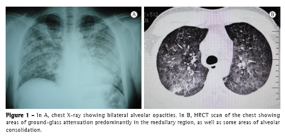
A chest X-ray revealed bilateral alveolar opacities (Figure 1A). A HRCT scan of the chest showed areas of ground-glass attenuation predominantly in the medullary region, with some areas of alveolar consolidation (Figure 1B). The diagnosis of pulmonary hemosiderosis was considered during the analysis of the HRCT scan of the chest. Fiberoptic bronchoscopy revealed no lesions. However, alveolar macrophages containing hemosiderin granules (siderophores) were found in the bronchoalveolar lavage fluid.

The investigation of iron-deficiency anemia showed positivity for antiendomysium and antigliadin antibodies. A colonoscopy revealed no abnormalities, and a biopsy of the ileal mucosa revealed lymphoid follicular hyperplasia. An esophagogastroscopy revealed mild erosive duodenitis. The biopsy findings were suggestive of inactive chronic non-atrophic (grade 1) gastritis, with negative results for Helicobacter pylori and an active chronic inflammatory process in the duodenal mucosa, with moderate villous flattening and areas of apparent intraepithelial lymphocytosis, consistent with the diagnosis of celiac disease.
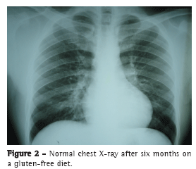
Within six months after the patient was started on a gluten-free diet, there was resolution of the pulmonary lesions and clinical improvement. At this writing, the patient was under follow-up in the department of pulmonology and was asymptomatic, chest X-ray findings and hemoglobin levels being normal (Figure 2).

Pulmonary hemosiderosis with celiac disease in a 29-year-old adult is rare, the number of cases being highest in children and adolescents.(4) In a study conducted in 2007, fifteen patients were under 20 years of age, and only five were over 20.(5) These data suggest that those diseases tend to occur in younger people.
The diagnosis of pulmonary hemosiderosis should be considered when the patient has the triad of recurrent hemoptysis, iron-deficiency anemia, and chest X-ray findings of infiltrate in the medullary region of the lung parenchyma. However, as in the present case, the clinical presentation can be atypical, without hemoptysis, which makes diagnosis difficult. In general, chest HRCT shows a ground-glass pattern, which predominantly occurs in the lower lung lobes and does not affect the periphery of the lung parenchyma. After other diseases have been ruled out by means of serological, microbiological, and radiological tests, bronchoalveolar lavage fluid showing hemosiderin-containing macrophages (siderophores) confirms the diagnosis. Subsequently, diseases or agents that are known to be related, among which is celiac disease, should be ruled out.(6) To that end, serology for antiendomysium and antigliadin antibodies should be performed even in the absence of gastrointestinal symptoms.(7)
It is important to confirm the presence of celiac disease, because treatment with a gluten-free diet is known to assist in the resolution of all symptoms and to result in radiological improvement in most individuals,(8) as seen in this clinical case. Long-term nutritional follow-up is also important; it has been reported that poor adherence to the diet can reactivate respiratory and gastrointestinal symptoms.(3)
In conclusion, the association between pulmonary hemosiderosis and celiac disease in adults is rare but should not be overlooked, given that the pulmonary disease has significant clinical impact. If left untreated, it can have a poor prognosis, with the development of pulmonary fibrosis and chronic airflow limitation. Patients with pulmonary hemosiderosis should be investigated for celiac disease even in the absence of gastrointestinal symptoms. The available evidence, albeit scarce, demonstrates that a gluten-free diet is the most effective treatment for achieving remission of symptoms, although the mechanism by which the clinical respiratory symptoms improve remains unknown. In some cases, the use of corticosteroids and immunosuppressants can be beneficial, constituting a therapeutic option.(3)
José Wellington Alves dos Santos,
Head of the Pulmonology Department,
Santa Maria University Hospital,
Federal University of Santa Maria,
Santa Maria, Brazil
Abdias Baptista de Mello Neto,
Resident in Pulmonology,
Santa Maria University Hospital,
Federal University of Santa Maria,
Santa Maria, Brazil
Roseane Cardoso Marchiori,
Professor of Pulmonology,
Federal University of Santa Maria,
Santa Maria, Brazil
Gustavo Trindade Michel,
Professor of Pulmonology,
Federal University of Santa Maria,
Santa Maria, Brazil
Ariovaldo Leal Fagundes,
Pulmonologist, Pulmonology Department of the Santa Maria University Hospital,
Federal University of Santa Maria,
Santa Maria, Brazil
Leonardo Gonçalves Marques Tagliari,
Medical Student,
Federal University of Santa Maria,
Santa Maria, Brazil
Tiago Cancian,
Medical Student,
Federal University of Santa Maria,
Santa Maria, BrazilReferencesNuesslein TG, Teig N, Rieger CH. Pulmonary haemosiderosis in infants and children. Paediatr Respir Rev. 2006;7(1):45-8. PMid:16473816. http://dx.doi.org/10.1016/j.prrv.2005.11.003
Lane DJ, Hamilton WS. Idiopathic steatorrhoea and idiopathic pulmonary haemosiderosis. Br Med J. 1971;2(5753):89-90. PMid:5551274 PMCid:1795538. http://dx.doi.org/10.1136/bmj.2.5753.89
Khemiri M, Ouederni M, Khaldi F, Barsaoui S. Screening for celiac disease in idiopathic pulmonary hemosiderosis. Gastroenterol Clin Biol. 2008;32(8-9):745-8. PMid:18603390. http://dx.doi.org/10.1016/j.gcb.2008.05.010
Malhotra P, Aggarwal R, Aggarwal AN, Jindal SK, Awasthi A, Radotra BD. Coeliac disease as a cause of unusually severe anaemia in a young man with idiopathic pulmonary haemosiderosis. Respir Med. 2005;99(4):451-3. PMid:15763451. http://dx.doi.org/10.1016/j.rmed.2004.09.007
Agarwal R, Aggarwal AN, Gupta D. Lane-Hamilton syndrome: simultaneous occurrence of coeliac disease and idiopathic pulmonary haemosiderosis. Intern Med J. 2007;37(1):65-7. PMid:17199848. http://dx.doi.org/10.1111/j.1445-5994.2006.01226.x
Ioachimescu OC, Sieber S, Kotch A. Idiopathic pulmonary haemosiderosis revisited. Eur Respir J. 2004;24(1):162-70. PMid:15293620. http://dx.doi.org/10.1183/09031936.04.00116302
Agarwal R, Aggarwal AN, Gupta D. Case 3-2007: a boy with respiratory insufficiency. N Engl J Med. 2007;356(22):2329; author reply 2330. http://dx.doi.org/10.1056/NEJMc070497
Sethi GR, Singhal KK, Puri AS, Mantan M. Benefit of gluten-free diet in idiopathic pulmonary hemosiderosis in association with celiac disease. Pediatr Pulmonol. 2011;46(3):302-5. PMid:20967850. http://dx.doi.org/10.1002/ppul.21357


